m-mode lv echovcardiography | m mode echocardiogram left ventricular dysfunction m-mode lv echovcardiography Assessment of LV function with M-mode or 2-dimensional (2-D) echocardiography (Figure 2A) can be performed in the parasternal long- and short-axis views by . iMaxTree. With a Maxi Dress. The dramatic proportions of a chunky knit sweater and a floor-sweeping dress instantly look high fashion. I love this combination .
0 · m mode echocardiogram left ventricular dysfunction
1 · m mode echocardiogram ejection fraction
2 · m mode echo cardiography
3 · echocardiogram left ventricular dysfunction
4 · 2d echocardiography m mode
1. Explore your overseas Card Member benefits. Before you even embark on your journey, it’s a good idea to explore your Card’s travel and international perks. Look into using .
M-mode was previously the dominating modality in echocardiography. Although it has now largely been replaced by 2D echocardiography, it is still used in . M-mode echocardiogram is commonly used to measure left ventricular dimensions and ejection fraction. Ejection fraction is indicative of .Assessment of LV function with M-mode or 2-dimensional (2-D) echocardiography (Figure 2A) can be performed in the parasternal long- and short-axis views by . The first and most commonly used echocardiography method of LVM estimation is the linear method, which uses end-diastolic linear measurements of the interventricular septum (IVSd), LV inferolateral wall .
o Derived from M-mode or linear 2D measurements Ejection Fraction (EF) (see below) is the predominant method for assessing global systolic function (see below) and is derived from the .
M-mode echocardiography provides a single line of information at a higher frame rate than can be obtained by two-dimensional echocardiography. This technique enhances accurate . M-mode echo in left ventricular dysfunction due to anthracycline related cardiomyopathy – annotated C is the systolic closure point of the mitral valve. CD segment represents systole.
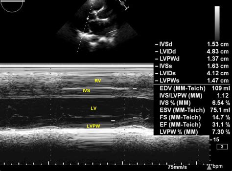
M-mode was previously the dominating modality in echocardiography. Although it has now largely been replaced by 2D echocardiography, it is still used in clinical practice. M-mode provides a one-dimensional view of all reflectors (i.e structures reflecting ultrasound waves) along one ultrasound line. Hence, the M-mode image displays all . M-mode echocardiogram is commonly used to measure left ventricular dimensions and ejection fraction. Ejection fraction is indicative of the left ventricular systolic function. In this case left ventricular systolic function is grossly depressed, with a left ventricular ejection fraction (EF) of only 31.1%.
m mode echocardiogram left ventricular dysfunction
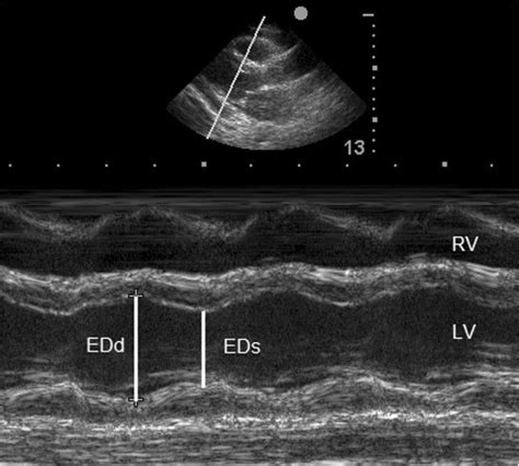
Assessment of LV function with M-mode or 2-dimensional (2-D) echocardiography (Figure 2A) can be performed in the parasternal long- and short-axis views by placing the calipers perpendicular to the ventricular long axis. Change in LV cavity dimensions during systole can be used to calculate LV fractional shortening and ejection fraction. The first and most commonly used echocardiography method of LVM estimation is the linear method, which uses end-diastolic linear measurements of the interventricular septum (IVSd), LV inferolateral wall thickness, and LV internal diameter derived from 2D-guided M-mode or direct 2D echocardiography. This method utilizes the Devereux and Reichek .o Derived from M-mode or linear 2D measurements Ejection Fraction (EF) (see below) is the predominant method for assessing global systolic function (see below) and is derived from the LV end-diastolic volume (LVEDV) and LV end-systolic volume (LVESV). Global Longitudinal Strain is a new parameter to assess LV systolic function.
M-mode echocardiography provides a single line of information at a higher frame rate than can be obtained by two-dimensional echocardiography. This technique enhances accurate determination of linear dimensions and improves quantitation of chamber size and wall thickness.
M-mode echo in left ventricular dysfunction due to anthracycline related cardiomyopathy – annotated C is the systolic closure point of the mitral valve. CD segment represents systole.

M-Mode. Evaluate wall contractility (wall thickness, systolic wall thickening, systolic wall motion) B -"bump" or "notch" of mitral valve indicates increased LVEDP ≥15 mm Hg. B "bump" or "notch" tricuspid valve indicates increased RVEDP ≥9 mm Hg. 10 reasons to use M-mode Echocardiography. Better understanding of LA hemodynamics. Better estimation of time intervals. Simple evaluation of dyssynchrony. Motion patterns of normal and abnormal structures. Identify high frequency motion. Rapidly moving structures such as the aortic valve and mitral valve, and endocardium have characteristic movements in M-mode. M-mode also has a great spatial resolution, which is useful for measuring ventricular dimensions in systole and diastole.M-mode was previously the dominating modality in echocardiography. Although it has now largely been replaced by 2D echocardiography, it is still used in clinical practice. M-mode provides a one-dimensional view of all reflectors (i.e structures reflecting ultrasound waves) along one ultrasound line. Hence, the M-mode image displays all .
M-mode echocardiogram is commonly used to measure left ventricular dimensions and ejection fraction. Ejection fraction is indicative of the left ventricular systolic function. In this case left ventricular systolic function is grossly depressed, with a left ventricular ejection fraction (EF) of only 31.1%.Assessment of LV function with M-mode or 2-dimensional (2-D) echocardiography (Figure 2A) can be performed in the parasternal long- and short-axis views by placing the calipers perpendicular to the ventricular long axis. Change in LV cavity dimensions during systole can be used to calculate LV fractional shortening and ejection fraction. The first and most commonly used echocardiography method of LVM estimation is the linear method, which uses end-diastolic linear measurements of the interventricular septum (IVSd), LV inferolateral wall thickness, and LV internal diameter derived from 2D-guided M-mode or direct 2D echocardiography. This method utilizes the Devereux and Reichek .o Derived from M-mode or linear 2D measurements Ejection Fraction (EF) (see below) is the predominant method for assessing global systolic function (see below) and is derived from the LV end-diastolic volume (LVEDV) and LV end-systolic volume (LVESV). Global Longitudinal Strain is a new parameter to assess LV systolic function.
M-mode echocardiography provides a single line of information at a higher frame rate than can be obtained by two-dimensional echocardiography. This technique enhances accurate determination of linear dimensions and improves quantitation of chamber size and wall thickness. M-mode echo in left ventricular dysfunction due to anthracycline related cardiomyopathy – annotated C is the systolic closure point of the mitral valve. CD segment represents systole.M-Mode. Evaluate wall contractility (wall thickness, systolic wall thickening, systolic wall motion) B -"bump" or "notch" of mitral valve indicates increased LVEDP ≥15 mm Hg. B "bump" or "notch" tricuspid valve indicates increased RVEDP ≥9 mm Hg. 10 reasons to use M-mode Echocardiography. Better understanding of LA hemodynamics. Better estimation of time intervals. Simple evaluation of dyssynchrony. Motion patterns of normal and abnormal structures. Identify high frequency motion.
prada cologne mercado libre

bolsos prada precio colombia
The distance between Netherlands and Malta is 2005 km. How do I travel from Netherlands to Malta without a car? The best way to get from Netherlands to Malta without a car is to train and bus and bus and ferry which takes 42h 56m and costs $260 - $430.
m-mode lv echovcardiography|m mode echocardiogram left ventricular dysfunction


